VDG-Nachrichten 04/2020
Training Aid for Aortic Stent Placement
By: Steve Anderson, Mayo Clinic, Division of Engineering, Rochester, MN 55905. Email: anderson.steven@mayo.edu. Claire Eggleston, Tyler King, Victoria Lundine, Ebenfalls Mayo Clinic, Division of Engineering, Rochester, MN 55905. Gustavo Oderich, M.D., Jill Colglazier, M.D., Raymond Shields M.D., Mayo Clinic, Division of Vascular and Endovascular, Rochester, MN 55905. Karen Marie Bjellum, Mayo Clinic, Division of Surgical Services, Rochester, MN 55905.
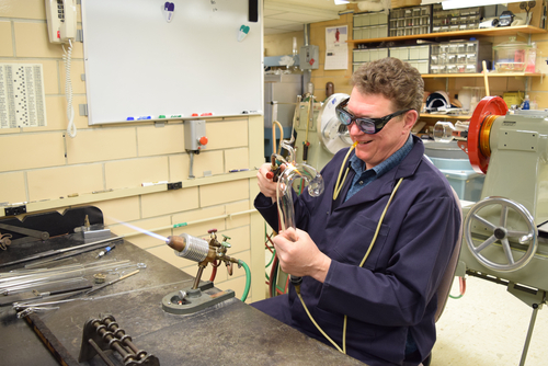
Aortic training model fabricated of borosilicate glass used in the education and training of fellows and residents in vascular surgery.
Background:
Aortic diseases represent a variety of conditions from clinically silent to acutely symptomatic, which affect virtually any part of the aorta. In-depth studies of aortic disease conducted increasingly over the past two decades have significantly enhanced our understanding that the aorta is an active living organ and not just a passive hollow conduit that transports blood.
Objective:
The aortic training model will be used in education and training of the fellows and residents in vascular surgery. The model is of the aorta in its entirety with its major arterial branches and can replicate aneurysmal disease of the aorta in its arch, descending thoracic aorta or abdominal aorta and iliac arteries. The fellows and residents will learn wire and catheter skills involved in endovascular procedures while using this model. They will be able to perform entire endovascular procedures that range from treating standard infrarenal abdominal aortic aneurysms, to more complex fenestrated and branched endografts to treat extent II throacoabdominal aortic aneurysms that involve cannulating the visceral arteries. This training model will provide a safe environment for the trainees to learn and practice these advanced endovascular skills. One of the biggest issues with this procedure is radiation and with this model students are able to practice without exposure to radiation and prior to being involved in the same procedure with a patient.
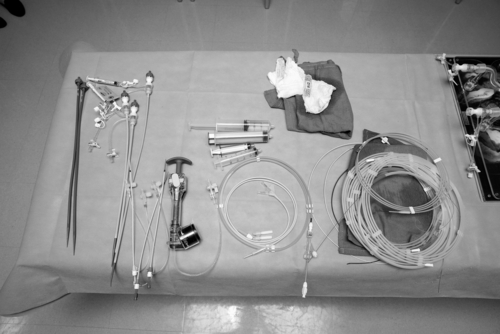
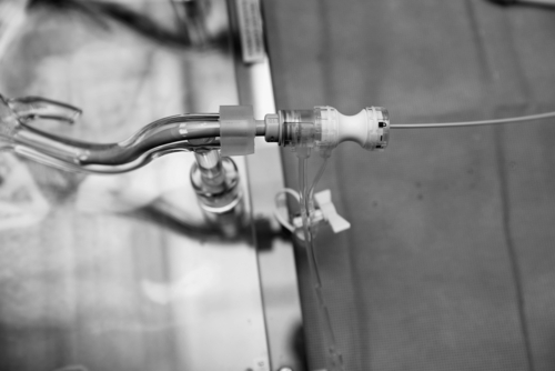
Instruments used in percutaneous placement of aortic endograft to treat abdominal aortic aneurysms. From left to right sheaths, balloon insufflator, aortic occlusion balloon, syringes, multiple wires.
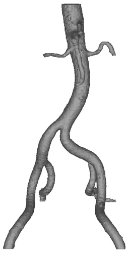
Figure 1 shows the abdominal aorta from a clinical CTA in the Mimics software which was used to measure the diameter of the vessels. The center line from the model was used in creating the SOLIDWORKS model in Figure 2.
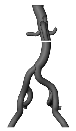
Figure 2 contains an image of the SOLIDWORKS model that was created using the center line and vessel diameters from the Mimics model in Figure 1.
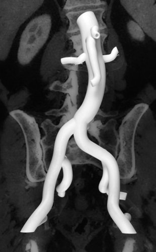
Figure 3 is an image of the 3D printed model which was printed on the Fortus 400 using the Solidworks models in Figure 2.
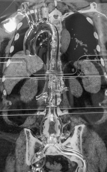
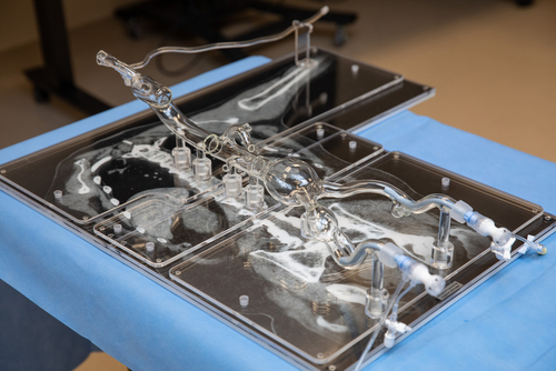
Image 4: The glass model portion of the training aid is fabricated using borosilicate glass tubing. The arch and trunk were made with 28 mm o.d. x 22 mm i.d. and the Iliac uses 16 mm o.d. x 11 mm i.d. The glass model is connected to the base by using 12 mm o.d. rod as stand offs.
Image 5: Glass model of the aorta from the aortic valve to the common femoral arteries with the left brachial artery and bilateral common femoral arteries as access sites for percutaneous procedures. 18 French sheaths are seen in the common femoral arteries bilaterally.

The image on the left shows sheath access into the common femoral artery. 18 French sheaths with catheter coming out of it. The image on the right shows delivery of the aortic endograft through the right groin access sheath up to the level of the bilateral renal arteries. The aortic endograft has not been deployed yet.
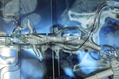
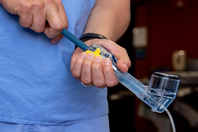
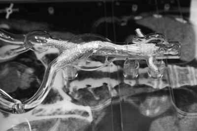
Image 9 shows the deployment of bilateral renal artery stents with a deployed aortic endograft in place. The image on the right shows the balloon insufflation of renal artery balloon expandable stent graft to deploy the stent graft.
Image 10: The aortic stent graft is completed with two bilateral renal artery stents. Through the use of the snorkel technique, the two bilateral renal stents are placed and deployed along with an aortic endograft. Without the bilateral renal stents the aortic endograft would restrict blood flow to the renal arteries which supply blood to the kidneys.
Conclusion
The Training Aid for Aortic Stent Placement will allow fellows and residents to practice an intricate procedure without being exposed to radiation or them potentially injuring a patient, both of these things would be possibilities in the operating room. The glass blown models allow these students to better understand the most common types of aneurysms as well as the behaviors and tendencies of the devices they will be using in surgery. Fluoroscopy is used in surgery which allows the surgeon to see the stent and anatomy of the patient in 2D as opposed to 3D with the glass models.

This article was presented as a poster at the ASGS Symposium, held in Corning, NY, USA in June 2019. As a part of the closer cooperation of the VDG and the ASGS we were happy to receive this article for reprint. Thank you very much for your cooperation.
Read the article in german
VDG-Nachrichten
Herausgeber
Verband Deutscher Glasbläser e.V., Karlstr. 7, D-48268 Greven
Erscheinungsweise
vierteljährlich im März, Juni, September und Dezember (Sonderausgabe nach Bedarf)
Format
210 x 297 mm (DIN A 4)
Bezugspreis
Der Mitgliedsbeitrag beinhaltet den Bezug der VDG-Nachrichten; Bezugspreis für Nichtmitglieder: 13,50 €.
Druck
Karl Elser Druck GmbH, Mühlhacker / E-Mail: info@elserdruck.de
Kontakt, Anfragen und Anzeigenschaltung:
Redaktion des VDG e.V.
c/o Eberhard Karls Universität Tübungen
CZI Glasbläserei
Karin Rein und Thomas Nieß
Auf der Morgenstelle 6
72076 Tübingen